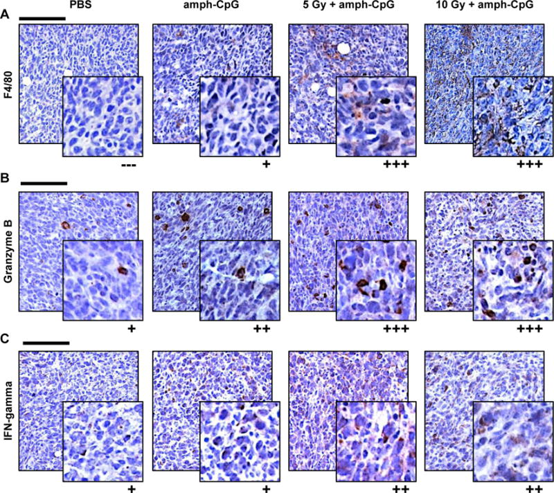
Figure 4.

Up-regulation of immune activity evident within TUBO tumors 48 hrs following radiation-enhanced delivery of amph-CpG. A) The increase in circulating monocytes seen after radiation-enhanced delivery coincided with an observed macrophage increase in tumor sections by IHC (brown = F4/80, macrophages). B) CD8+ T lymphocytes in tumor sections displayed increased Granzyme B immunoreactivity (brown) after IR. C) NK cell activity (brown = IFNγ) appeared up-regulated in tumor sections following IR + amph-CpG. In all IHC images, purple = hematoxylin, nuclei. Scale bar = 100 μm. Inset = 4X zoom. Qualitative scoring: (−−−) negative, (+) faint staining of some cells, (++) moderate staining of many cells, (+++) intense staining of many cells. Representative images shown of n = 3 tumors per data set stained.
