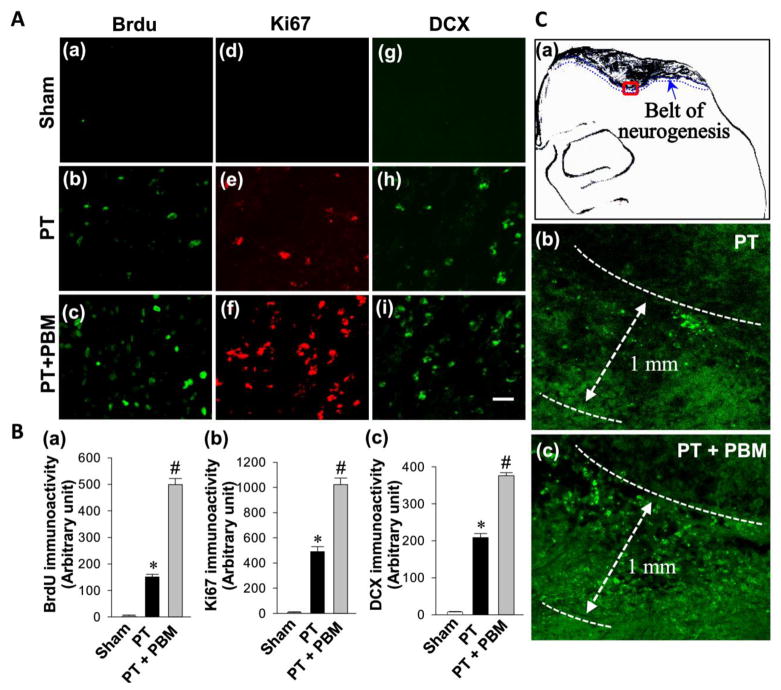
Fig. 5. LLI improves neurogenesis in the peri-infarct cortical region on day 14 after PT stroke.
(A) Representative confocal microscopy images of BrdU (a–c), Ki67 (d–f) and DCX (g–i) were taken from the cortical peri-infarct region on day 14 after PT stroke. (B) The immunoactivity associated with BrdU, Ki67 and DCX in each group were further quantified and shown in arbitrary unit (a, b and c respectively). (C) The increased neurogenesis was restricted to a regions (around 1.00 mm wide belt) beneath the infarct zone surface with the representative confocal microscopy of Brdu staining (a–c). All data are expressed as mean ± SE (n = 4–5). *P < 0.05 versus sham, #P < 0.05 versus PT. Scale bar = 20 μm.

