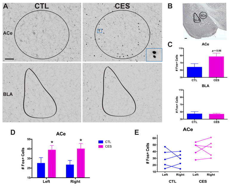
Figure 3.
The central nucleus of the amygdala (ACe) is aberrantly activated during social play in CES adolescent rats. (A) Representative images of Fos+ cells in the counted regions, including ACe (top) and BLA (bottom), in CTL and CES adolescent rats sacrificed 90 min. following social play (Cohort 2). Scale bar = 100 μm. (B) Low-magnification image of the brain regions where Fos+ cells were counted, including the ACe and BLA. Scale bar = 200 μm. (C) Although the pleasurable experience of play induced c-Fos activation in multiple brain regions in both groups of rats, a key region that distinguished CES from CTL was the ACe: Social play activated a greater number of cells in the ACe of CES rats compared to controls (top), whereas Fos activation in the neighboring BLA did not distinguish between the groups (bottom). (D) When analyzed separately, both the right and left hemispheres of the ACe showed an increase of Fos+ cells in CES rats following social play. (E) Within individual rats, the number of Fos+ cells activated on the right vs. left hemisphere of the ACe did not significantly differ. Values are expressed as mean ± SEM (n = 5 rats per group; *p = 0.05).

