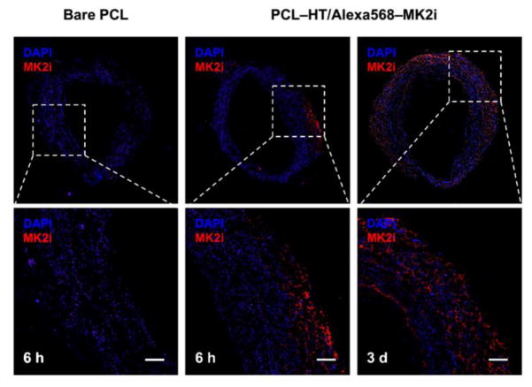
Figure 7. MK2i peptide delivery of PCL–HT/MK2i to human vein tissue.
Representative fluorescence images of the HSVs wrapped with bare PCL and PCL–HT/MK2i sheaths for 6 h and 3 days. MK2i peptide was labelled with Alexa 568 (red) before loading on the surface of PCL–HT sheath. The cross-sectioned HSVs were stained with DAPI (blue). Scale bars indicate 200 μm.

