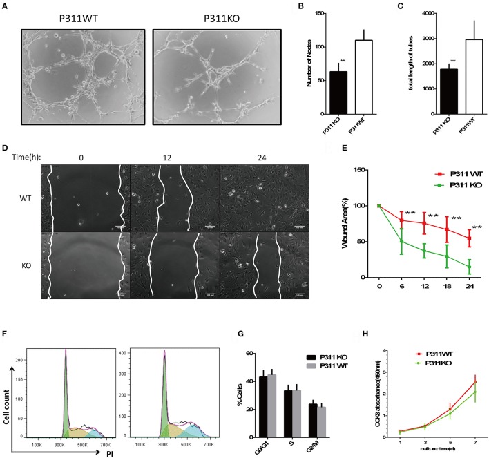
Figure 2.
Effect of P311 deficiency on endothelial cell function in vitro. (A) Representative images of tube formation in Matrigel. Scar bar = 100 μm. (B,C) Quantitation of tube formation (n = 6 per group). (B) Number of nobes. (C) Total length of tubes. (D) Representative images of recovery of scratched areas by cell migration at 12 and 24 h (n = 6 per group). The white line stands for the leading edge of migration cells. Scale bars = 100 μm. (E) Quantitation of the wound area (n = 6 per group). (F) Representative flow cytometry cell cycle of PI. (G) Quantitation data of flow cytometry cell cycle (n = 5 per group). (H) CCK8 assay was performed to assess the effect of P311 on the proliferation (n = 6 per group). Data were expressed as means ± SD. **P < 0.01, P311 WT vs. KO. All experiments were repeated for three times with reproducible results.

