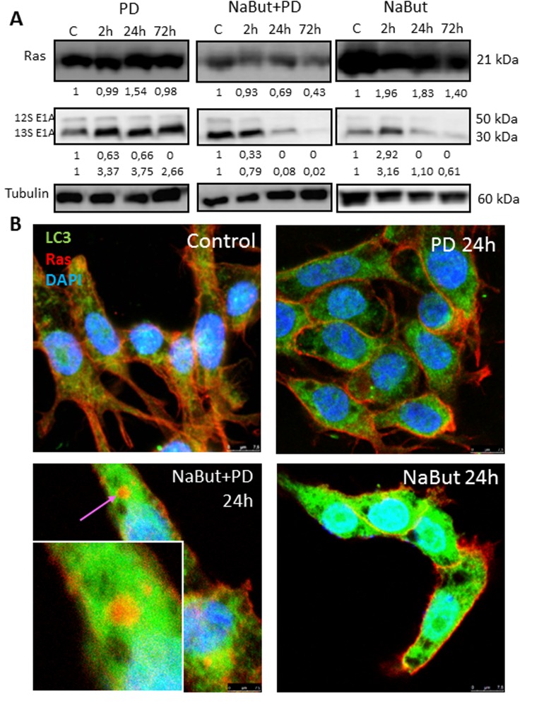
Figure 9. MEK/ERK suppression in senescent ERas cells changes balance of E1A and Ha-Ras oncoproteins and leads to relocalization of Ras from the plasma membrane into the cytoplasm.
(A) Western-blotting analysis of Ras and E1A (12S, 13S) expression. Cells were exposed to inhibitors and processed to Western-blotting in 12% gel. Numbers below present densitometry of bands. (B) Immunofluorescent images show changes of Ras (red) and LC3 (green) localization in senescent cells with suppressed MEK/ERK. Cells were treated with inhibitors for the indicated time, then fixed and stained with antibodies against pan-Ras and pan-LC3. Square indicates a magnified region showing Ras in the cytoplasm colocalized with LC3 in senescent PD-treated cell. Nuclei stained with DAPI (blue). Scale bars: 25 μm.

