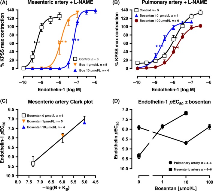
Figure 1.

Average single exposure concentration‐contraction curves to endothelin‐1 in rat (A) mesenteric artery (n = 15) and (B) pulmonary artery (n = 15), pretreated with L‐NAME 100 μmol L−1, in the absence Control, (0 μmol L−1) or presence of bosentan 1, 10 or 100 μmol L−1. Data are expressed as % KPSS maximum contraction (y axis). (C) Clark plot display for the relationship in the rat mesenteric artery between the endothelin‐1 pEC 50 values (y axis; −log M) and −log(B + KB) where B is concentration of bosentan (0, 1, or 10 μmol L−1) and KB is the global‐fitted dissociation constant. The error bars are ± 2 SEM of the difference between the nonlinear regression‐fitted pEC 50 values for endothelin‐1 and the pEC 50 values fitted for the individual artery for each concentration of bosentan (B). (D) The pEC 50 values for the endothelin‐1 curves in (A) and (B) are plotted on the y axis against the bosentan concentration (x axis) for each artery type. Vertical error bars in (A, B, and D) are ± 1 SEM (those not shown are contained within the symbol). Horizontal error bars (A‐B) represent the EC 50 ± 1 SEM. n, number of arteries isolated from separate animals. *P < .05, pEC 50 values compared with respective control (0 μmol L−1) pEC 50 values. Variations in n are due to violation of predetermined criteria: mesenteric arteries that contracted to KPSS with <3 mN force or pulmonary arteries that contracted to KPSS with <1 mN force
