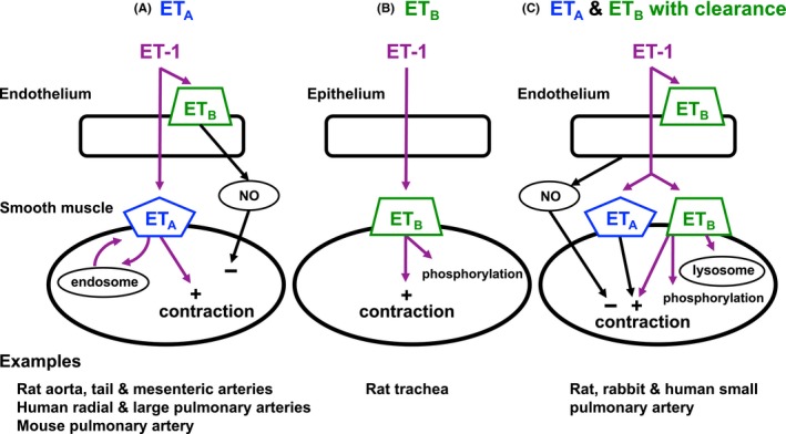
Figure 9.

Schematic diagram of the location and function of ETA and ETB receptors in 3 tissue assays. (A) ETA receptors located on vascular smooth muscle mediate contraction. ETA receptors are internalized and recycled slowly through endosomes.36 (B) ETB receptors located on smooth muscle cells mediate contraction and are rapidly removed by phosphorylation.36 (C) ETB receptors located on smooth muscle cells bind endothelin‐1 and clear endothelin‐1 from the environment through lysosomal metabolism. The remaining endothelin‐1 binds to ETA and ETB receptors on smooth muscle to mediate contraction before being recycled by endosomes or destroyed by phosphorylation, respectively. In (A) and (C), ETB receptors on the endothelium mediate release of NO that transiently relaxes smooth muscle. Examples of the species and tissues assumed to have these particular receptor profiles are given below each panel. ET‐1, endothelin‐1. NO, nitric oxide
