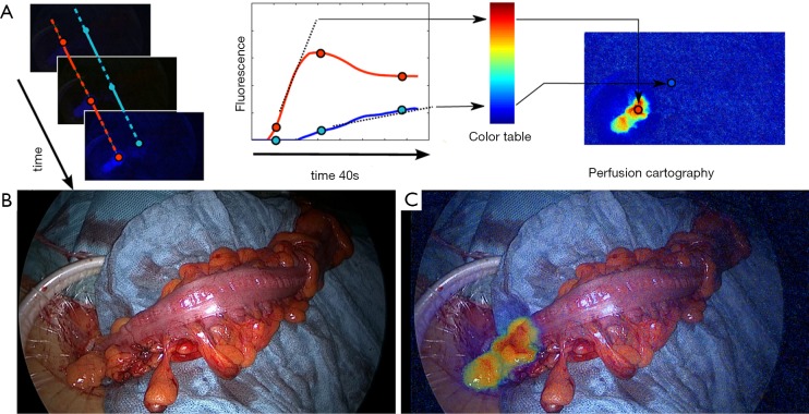
Figure 1.
Clinical application of the fluorescence-based enhanced reality to evaluate bowel perfusion. (A) The concept of FLER (fluorescence-based enhanced reality): after the i.v. administration of the Indocyanine Green, the fluorescence signal is analysed during 40 seconds by a specific software (VR-PERFUSION, IRCAD; France). The slope of the fluorescence time-to-peak is computed pixel-by-pixel and is converted into a color code to generate a virtual perfusion cartography. The resultant cartography is overlapped on real-time images [(B) white light images of the proximal resection during a sigmoidectomy] using a video-mixer [(C) augmented reality view of the proximal resection site].

