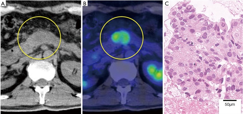
Figure 2.
PET-CT findings and microscopic findings of the pancreatic nodule. (A) PET-CT shows a 2.5-cm nodule in the pancreatic body. (B) Accumulation of FDG is detected in the nodule. (C) The nodule was diagnosed as a pancreatic metastasis of lung cancer using EUS-FNA biopsy. PET-CT, positron emission tomography-computed tomography; FDG, fluorine-18-deoxyglucose; EUS-FNA, endoscopic ultrasound-guided fine-needle aspiration.

