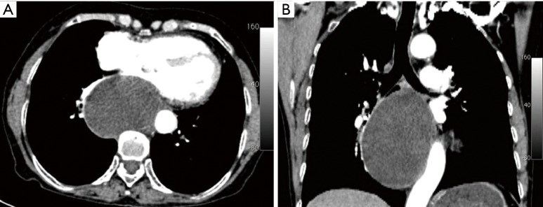
Figure 1.
Contrast-enhanced chest computed tomography (CT) revealed a large cystic mass in the posterior mediastinum in contact with the left main bronchial wall and the descending thoracic aorta. The mass was homogeneous, had regular borders and low density, and showed no enhancement after administration of contrast medium.

