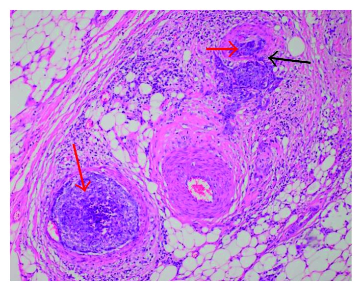
Figure 4.

Histological representation of metastasis in the left submandibular gland, showing basaloid cells, compatible with the cellular pattern of the primary site, inside the vessels, provoking tumor thrombi (red arrows). Presence of vascular invasion behavior, indicating probable hematogenous origin of metastases (black arrow).
