
Figure 4.
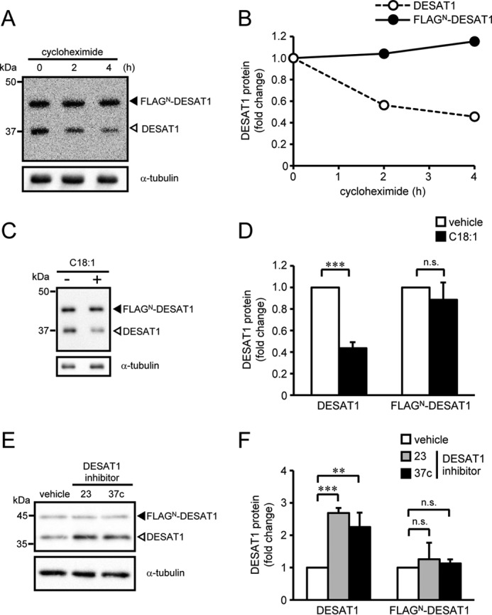
Effect of an N-terminal FLAG tag on DESAT1 expression. S2 cells expressing FLAGN-DESAT1 were treated with cycloheximide (100 μg/ml) for 0, 2, and 4 h, and the amounts of DESAT1 and α-tubulin protein were detected with anti-DESAT1 antibody and anti-α-tubulin antibody, respectively (A and B). S2 cells expressing FLAGN-DESAT1 were treated with C18:1 (100 μm) for 6 h (C and D) or DESAT1 inhibitor 23 or 37c (1 μm) for 16 h (E and F), and the amounts of DESAT1 and α-tubulin protein were detected with specific antibodies. Band intensities were determined by ImageJ software, and levels of DESAT1 proteins are shown relative to the amount of DESAT1 protein in cells not treated with cycloheximide (B) or vehicle-treated cells (D and F). Filled arrowhead, FLAGN-DESAT1; open arrowhead, endogenous DESAT1. Mean ± S.D. (n = 3). **, p < 0.01; ***, p < 0.001; n.s., not significant.