Figure 7.
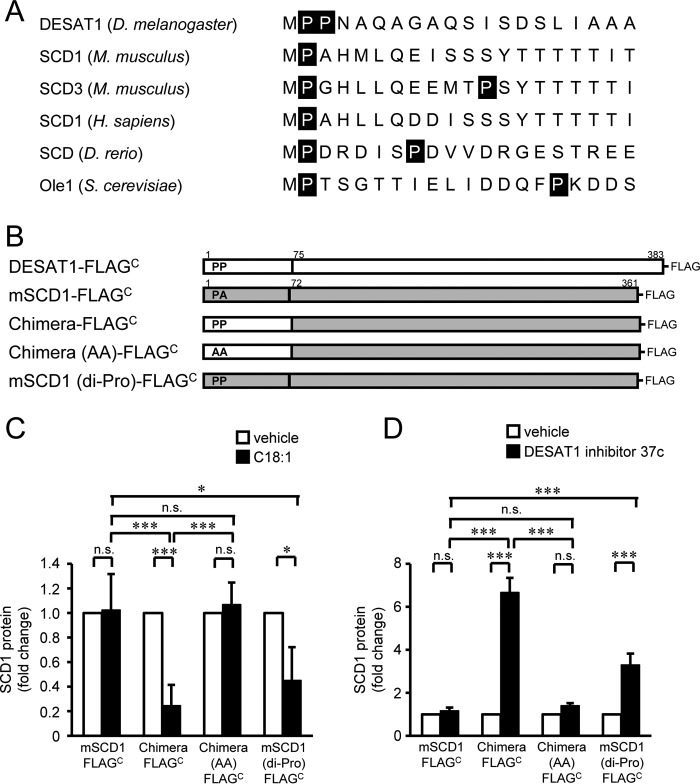
Role of the di-proline motif in degradation of Δ9-desaturase. N-terminal amino acid sequences of Δ9-desaturases were compared (D. melanogaster DESAT1 (NP_652731), Mus musculus SCD1 (NP_033153), M. musculus SCD3 (NP_077770), Homo sapiens SCD1 (NP_005054), Danio rerio stearoyl-CoA desaturase (AAO25582), and S. cerevisiae Ole1 (CAA96757)) (A). Schematic illustration of DESAT1, mouse SCD1, and constructed mutants is shown (B). S2 cells expressing mouse SCD1-FLAGC, chimera-FLAGC, chimera (AA)-FLAGC, and SCD1 (di-Pro)-FLAGC were treated with C18:1 (100 μm) for 6 h (C) or DESAT1 inhibitor 37c (1 μm) for 16 h (D), and the amounts of endogenous DESAT1, FLAG-tagged exogenous Δ9-desaturases, and α-tubulin protein were detected with anti-DESAT1 antibody, anti-FLAG antibody, and anti-α-tubulin antibody, respectively. Band intensities were determined by ImageJ software, and levels of SCD1 proteins are shown relative to the amount of SCD1 protein in vehicle-treated cells (C and D). Mean ± S.D. (n = 3). *, p < 0.05; ***, p < 0.001; n.s., not significant.

