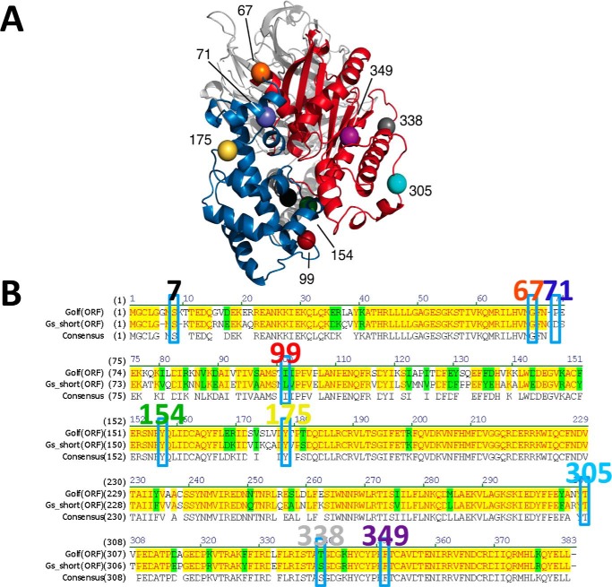
Figure 1.
A, location of the insertion points of six selected probe positions resolved in the inactive/closed (PDB code 1AZT) crystal structure of Gs are shown at positions 99 (red), 154 (green), 175 (yellow), 305 (light blue), 338 (gray), and 349 (purple). The α-helical and Ras-like domains of Gα are in blue and red, respectively, whereas Gβ and Gγ are in light gray and dark gray. B, amino acid sequence alignment between Golf and Gs short. Identical and homologous residues are highlighted in yellow and green, respectively. Insertion positions for Gs as well as Golf are enclosed by rectangles, and the residue numbers for Gs are shown above.

