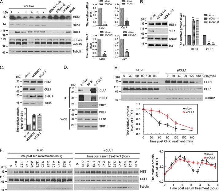
Figure 2.
CUL1 regulated HES1 stability and oscillation. A, Cullin family screening by siRNA. F9 cells were transfected with 50 nm Luc, RBX1, or Cullins siRNAs as indicated for 48 h, and the cells were harvested for Western blotting. Right panel, the mRNA level of CUL2, CUL3, CUL5, and CUL7 was examined using quantitative real-time PCR. The error bars represent S.D. from three independent experiments. The statistical difference was measured by paired two-sided Student's t test (**, p < 0.01). B, CUL1 regulated HES1 stability. F9 cells were transfected with 50 nm Luc or CUL1 (CUL1-1 and CUL1-2) siRNAs for 48 h, and the cells were harvested for Western blotting. The quantification of protein levels of HES1 and CUL1 were measured as in Fig. 1A. The error bars represent S.D. from three independent experiments. C, SCF is the major E3 ligase involved in HES1 degradation among CRL E3 ligases. F9 cells were transfected with 50 nm Luc and CUL1 siRNAs for 48 h or treated with 0.5 μm MLN4924 for 12 h, and cells were harvested for Western blotting. The quantification of protein levels of HES1 was measured as in Fig. 1A. The error bars represent S.D. from three independent experiments. The statistical difference was measured as in Fig. 1A. D, SCF complex interacted with HES1. F9 cells were treated with 20 μm MG132 for 3 h, the cells were harvested and analyzed by co-immunoprecipitation (IP) and Western blotting using antibodies against HES1, CUL1, and SKP1. Rabbit IgG (normal rabbit serum, NRS) was taken as negative control. Immunoblots of whole-cell extracts (WCE) are shown at the bottom. E, silencing of CUL1 resulted in stabilization of HES1. F9 cells were transfected with luciferase or CUL1 siRNAs for 45 h, and the cells were treated with CHX (100 μg/ml) for the indicated times. The protein levels of HES1, CUL1, and tubulin were determined by Western blotting. Bottom panel, the relative protein level of HES1 was quantified and normalized to tubulin. F, silencing of CUL1 disrupted HES1 oscillation. F9 cells were transfected with luciferase or CUL1 siRNAs for 24 h followed by serum-withdrawal starvation for 18.5 h. The cells were harvested at the indicated times once serum was supplemented. The protein level of HES1 was examined at the indicated times once serum was supplemented to the medium. Right panel, the quantification of relative protein level of HES1. The experiment was biologically repeated at least five times.

