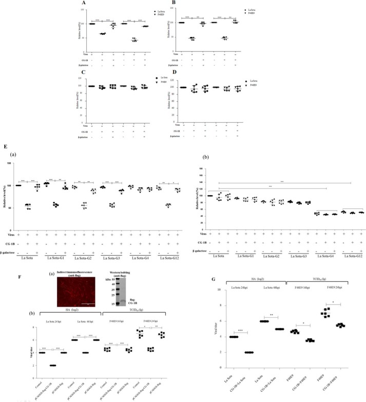
Figure 8.
Reduction of NDV adsorption and replication in DF-1 cells by CG-1B. A–C, adsorption assay. A, DF-1 cells were infected with NDV F48E9 or La Sota at m.o.i. 1 in the presence or absence of 7 μm CG-1B during 1 h at 4 °C. B, F48E9 and La Sota were pre-incubated with 7 μm CG-1B for 1 h at 37 °C before inoculation into DF-1 cells at 4 °C for 1 h. C, DF-1 cells were pre-incubated with 7 μm CG-1B for 1 h at 37 °C. Cells were washed three times with PBS and incubated with F48E9 or La Sota for 1 h at 4 °C. D, internalization assays. DF-1 cells were inoculated with NDV F48E9 and La Sota at m.o.i. 1 at 4 °C for 1 h. Cells were washed and incubated at 37 °C for 1 h supplemented with 7 μm CG-1B or medium. Un-internalized viruses were inactivated with citrate buffer. E, panel a, effect of CG-1B on the adsorption of HN mutants. DF-1 cells were inoculated with La Sota, La Sota-G1, -G2, -G3, -G4, and -G12 in the presence or absence of 7 μm CG-1B during 1 h at 4 °C. Panel b, effect of CG-1B on the internalization of HN mutants. DF-1 cells were inoculated with La Sota, La Sota-G1, -G2, -G3, -G4, and -G12 at 4 °C for 1 h. Then, cells were washed and incubated at 37 °C for 1 h supplemented with 7 μm CG-1B or medium. Un-internalized viruses were inactivated with citrate buffer. In all cases, β-galactose was used as an antagonist. Viral RNA was quantified by real-time RT-PCR. Data were normalized with expression of 18S rRNA gene, and the copy number of viral RNA was calculated relative to that of the La Sota group (×100%). F and G, CG-1B reduced replication of NDV in DF-1 cells. F, panel a, expression of FLAG-tagged CG-1B protein in DF-1 cells was identified through indirect immunofluorescence and Western blotting using anti-FLAG antibody. Panel b, quantification of NDV produced from pCAGGS-FLAG-CG-1B-transfected, pCAGGS-FLAG-transfected, and untransfected DF-1 cells after infection with F48E9 and La Sota. G, quantification of NDV produced from F48E9- and La Sota-infected DF-1 cells treated with CG-1B after adsorption. Viral titer in the culture supernatants of F48E9-infected cells was determined at 16 and 24 hpi by the Reed-Muench method in DF-1 cells, whereas viral titer in the culture supernatants of La-Sota-infected cells was determined at 24 and 48 hpi by HA assay. Virus titer assay was performed three times for each condition and compared using Student's t test (*, p < 0.05; **, p < 0.01; ***, p < 0.001). The bars represent means and S.D.

