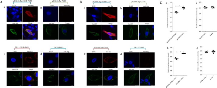
Figure 9.
Intracellular distribution and cell-surface expression of HN protein. A and B, intracellular distribution of CG-1B and NDV HN glycoprotein. Distribution of CG-1B and HN glycoprotein was detected in DF-1 cells transfected with pCAGGS-FLAG-CG-1B (panel a) and pCAGGS-FLAG (panel b), and DF-1 cells treated with 7 μm CG-1B after incubation (panel c). Untreated DF-1 cells were used as controls (panel d). NDV-infected cells were fixed at 24 h after infection and then subjected to indirect immunofluorescence to detect CG-1B and NDV HN glycoprotein using rabbit anti-FLAG/His antibody (red) and chicken anti-NDV HN antibody (green). The nuclei were stained with DAPI (blue) in the merged images. The triple-stained cells were observed by Leica SP2 laser confocal microscopy. C, cell-surface expression of HN was determined by flow cytometry. DF-1 cells transfected with pDsRed-CG-1B or pDsRed2-N1 were infected with NDV F48E9 (panel a) and La Sota (panel b) at m.o.i. 0.1. Surface expression of the HN protein in red fluorescence-positive DF-1 cells was assessed by flow cytometry at 24 hpi with an NDV HN-specific monoclonal antibody followed by a FITC-conjugated secondary antibody. In addition, surface expression of the HN protein in F48E9-infected (panel c) and La Sota-infected (panel d) DF-1 cells treated with 7 μm CG-1B after incubation was also detected. Surface immunofluorescence was quantified by fluorescence-activated cell sorter analysis with a Cytomics FC 500 flow cytometer.

