Figure 4.
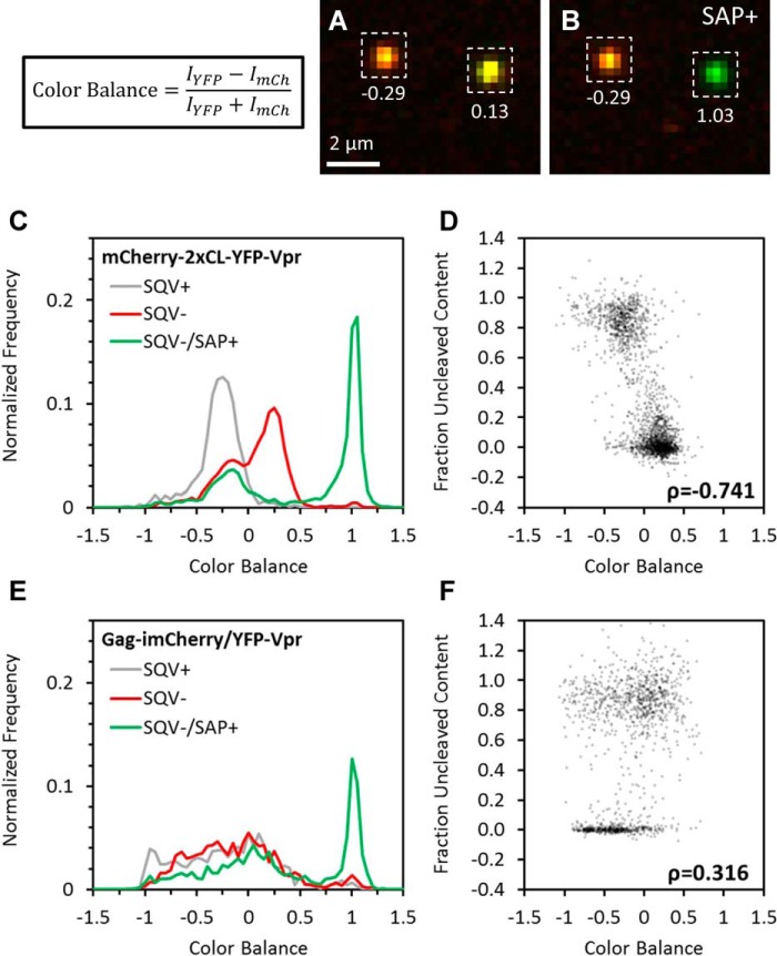
Single-particle color balance as an indicator of HIV-1 protease activity. HXB2 pseudoviruses labeled with either the bifunctional marker or Gag-imCherry/YFP-Vpr (data not shown) and treated with 500 nm SQV or left untreated were immobilized and imaged at high signal-to-noise before and after the addition of saponin. Single-particle color balance (see “Experimental procedures”) was measured using local background-corrected sum intensities of particles in the YFP and mCherry channels. A and B, representative images and measured color balance of particles labeled with bifunctional markers before (A) and after (B) addition of saponin. C and E, single-particle color balance distributions of HXB2 pseudovirus labeled with the bifunctional marker (C) or Gag-imCherry/YFP-Vpr (E) produced in the presence (gray) or absence of saquinavir before (red) and after (green) addition of saponin. D and F, scatterplot of single-particle color balance before saponin treatment and uncleaved mCherry content fraction of SQV− virus labeled with the bifunctional marker (D) or Gag-imCherry/YFP-Vpr (F). Color balance strongly (Pearson correlation, ρ = −0.741) and weakly (Pearson correlation, ρ = 0.316) correlated with uncleaved mCherry content for bifunctional labeling and Gag-imCherry/YFP-Vpr, respectively.

