Figure 5.
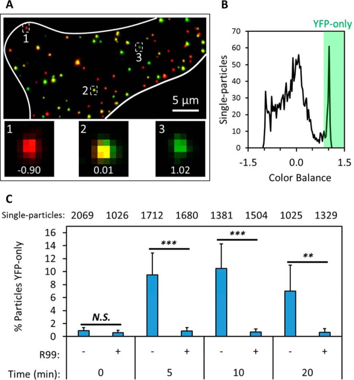
Detection of post-fusion viral cores in fixed cells. ASLV pseudoviruses labeled with the bifunctional marker were prebound to TVA950-expressing CV1 cells in the cold, and virus fusion was initiated by shifting to 37 °C for a short interval, after which cells were immediately fixed and imaged. A, maximum intensity projection of a fixed cell (white boundary) in which the virus was allowed to enter and fuse for 5 min. Insets highlight selected virus particles and their color balance, calculated from local background-corrected single-particle YFP and mCherry intensities. B, distribution of single-particle color balance at the 5-min time point. Post-fusion cores (YFP-only) are detected as puncta with color balance > 0.85. C, fraction of YFP-only particles per total particles in a field of view was measured for five to seven fields for each incubation interval. Parallel samples maintained in the presence of 50 μg/ml of the fusion inhibitor R99 were examined as negative controls. The total number of particles examined for each condition is listed on top. Error bars indicate standard deviation among the fields imaged for each condition. **, p < 0.01; ***, p < 0.001; N.S., not significant.

