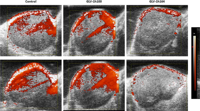
Figure 6. Doppler ultrasonography measurement of vascular flow in controls, GLV-1h100-, and GLV-1h164-infected tumors in vivo.

Two tumors from each treatment group were separately treated for 3 weeks and imaged with Doppler ultrasonography. The Doppler-derived relative tissue perfusion image was displayed in a “hot-iron” color scale superimposed on a gray-scale B-mode image, with lack of the hot-iron coloration corresponding to lower perfusion. Equal-size tumors were selected to avoid tumor size as confounding factor for vascular flow.
