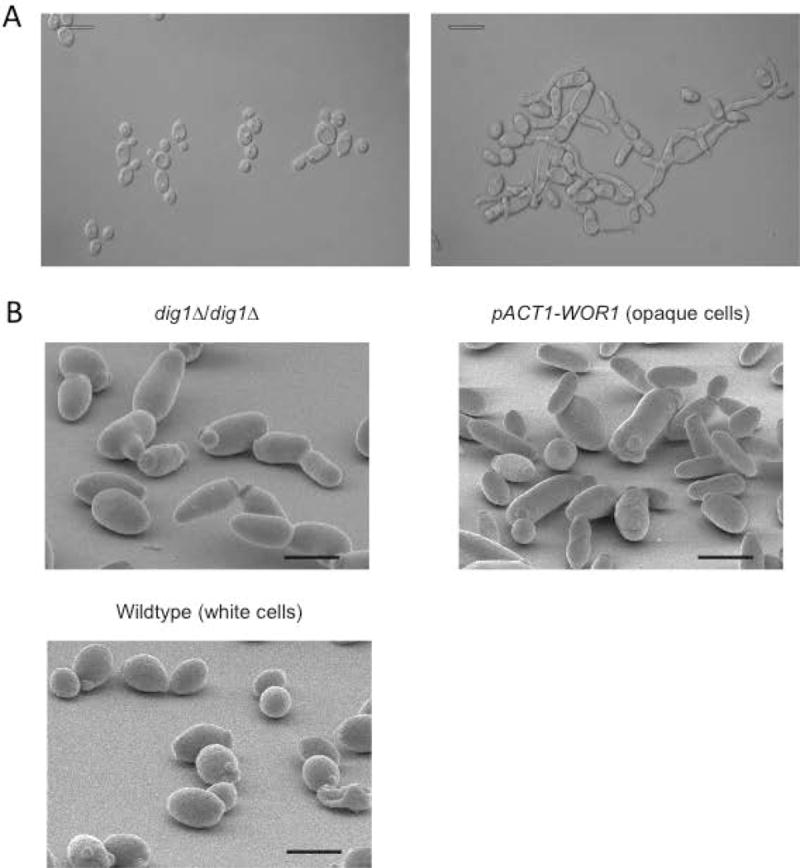
Figure 1. Imaging.

(A) Overnight cultures of wild-type SN148 (right panel) or HJR8 dig1Δ/dig1Δ (left panel) imaged by Differential Interference Contrast (DIC) microscopy show mixed morphology of the mutant cells. Scale bar 10 μm.
(B) C. albicans strain HJR8 dig1Δ/dig1Δ was inoculated onto YPD agar and incubated at 30 °C for 24 hours before processing for scanning electron microscopy; comparison of the dig1Δ strain with opaque (pACT1-WOR1) and white phase cells. Scale bar 15 μm.
