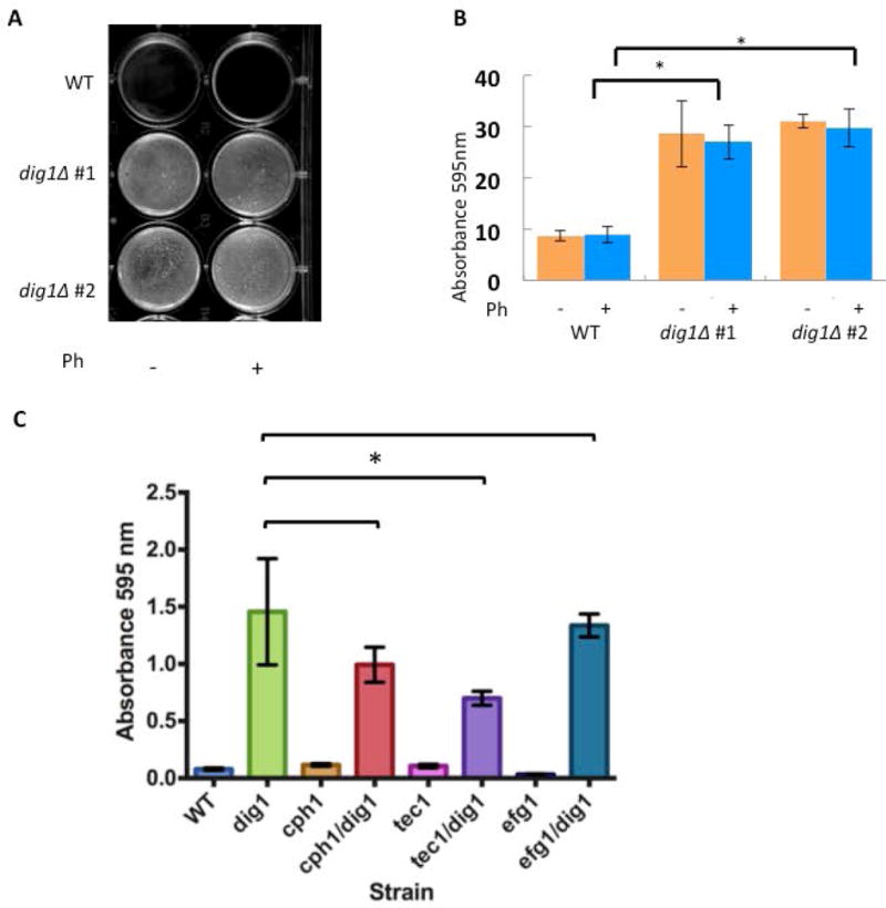
Figure 3. Enhanced biofilm formation in dig1Δ mutants.

(A) Representative image of biofilm formation in two independent MTLa dig1Δ mutants (CAY3570 and CAY3571) and the wild type control (RBY1132). Cells were grown as pheromone-stimulated biofilms in SCD media for 48 hours at room temperature without shaking. Ph = 10 μM α-pheromone. Reports (16, 26) both describe increased sexual biofilm in the SC5314 homozygote MTLa control compared to the regular biofilm. We believe the lack of difference in our data might be due to the fact we are using a different wild type control.
(B) Cells from strains CAY3570 and CAY3571were washed with PBS and adherence quantified by crystal violet staining. *= p <.01, Student’s t-test. error bars = standard deviation. n= 3 biological replicates.
(C) To determine the role of each transcription factor (Cph1p, Tec1p, Efg1p) in the formation of dig1∆/dig1∆-enhanced biofilms, wild type, single and double homozygous deletion mutants were challenged to form biofilms in the absence of pheromone. Tec1p is the major contributor to the formation of enhanced dig1∆/dig1∆ biofilms. All strains are MTLa/α. Cells were incubated in 6 well plates in SCD media for 24–48 hours at room temperature without shaking, washed with PBS and stained with crystal violet. Absorbance at 595 nm was recorded. Student’s unpaired t-test. Standard deviation indicated by error bars, n=3. *=p < 0.05.
