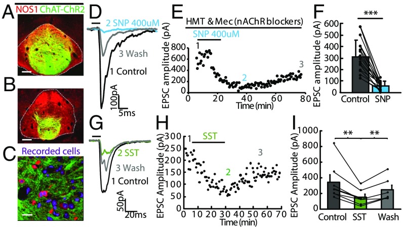
Fig. 4.
NO and SST inhibit evoked neurotransmitter release from axonal terminals of MHb neurons to the IPN. (A and B) Coronal sections from a ChAT-ChR2-EYFP mouse showing expression of NOS1 (red) in ChR2-EYFP+ terminals (green) in the IPN. (Scale bar: 100 µm.) (C) High-magnification view shows that ChR2-EYFP–labeled axonal terminals (green) encircle NOS1 immunopositive neurons (red) in the IPN. (Scale bar: 20 µm.) (D and E) Representative electrophysiological traces (D) and plot of EPSC amplitude versus time (E) show that application of the NO donor sodium nitroprusside (SNP) (400 µM) into the bath solution completely abolishes fast EPSCs evoked by brief blue light illumination (5 ms; horizontal bar) in the presence of nAChR blockers hexamethonium (HMT) and mecamylamine (Mec) and this blockade lasted for over 1 h. (F) Summary data show that SNP eliminates EPSCs (***P < 0.001, paired t test, n = 15 cells). Separate lines represent data from individual neurons. Control indicates EPSCs measured in nAChRs blockers. (G–I) Representative electrophysiological traces (G) and plot of EPSC amplitude versus time (H) show that application of SST (1 µM) reduces fast EPSCs evoked by brief blue light illumination (5 ms; horizontal bar) and this inhibition lasted for over 20 min in wash solution. (I) Summary data show that SST reduces EPSCs (**P < 0.005, paired t test, n = 6 cells). Separate lines represent data from individual neurons. Error bars indicate SEM.

