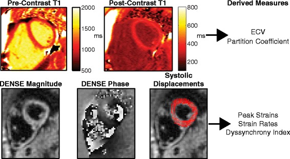
Fig. 1.

Example MOLLI T1 and DENSE images. Top row shows Pre- and Post-Contrast MOLLI T1 images for a representative patient. Bottom row shows DENSE magnitude and phase images for the same patient and same slice near end-systole, as well as the resulting displacement vectors at the same instant. The derived measures from each imaging sequence are listed for clarity
