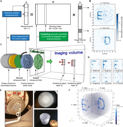
Fig. 6. Compressive 3D ultrasound imaging using a single sensor.

(A) Schematic sketch of the signal model involved in this type of compressive imaging. Each column of the observation matrix H contains the ultrasound pulse-echo signal that is associated with a pixel in 3D space, which is contained in the image vector v. By rotating the coding mask in front of the sensor, we obtain new measurements that can be stacked as additional entries in the measurement vector u and additional rows in H. (B) Result of solving part of the image vector v using an iterative least squares technique. The two images are mean projections of six pixels along the z dimension [individual z slices shown in (D)]. (C) Schematic overview of the complete imaging setup. A single sensor transmits a phase uniform ultrasound wave through a coding mask that enables the object information (two plastic letters “E” and “D”) to be compressed to a single measurement. Rotation of the mask enables additional measurements of the same object. (D) Reconstruction of the letter “E” in six adjacent z slices. A small tilt of the letter (from top left corner to bottom right corner) can be observed, demonstrating the potential 3D imaging capabilities of the proposed device. (E) Image showing the two 3D-printed letters and the plastic coding mask with a rubber band for rotating the mask over the sensor. The two right-hand panels show close-ups of the plastic coding mask. (F) 3D rendering of the complete reconstructed image vector v, obtained by BPDN. The images shown in (B) and (D) were obtained using 72 evenly spaced mask rotations, and the full 3D image in (F) was obtained using only 50 evenly spaced rotations to reduce the total matrix size.
