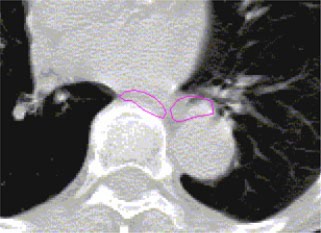
Figure 3.

(Color) A transverse plane showing the purported esophagus outlined by two different dosimetrists. Observe that there is no overlap between the esophagus as delineated by the two dosimetrists.

(Color) A transverse plane showing the purported esophagus outlined by two different dosimetrists. Observe that there is no overlap between the esophagus as delineated by the two dosimetrists.