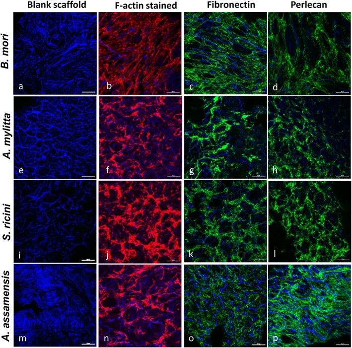
Figure 2.

Human skeletal muscle myoblasts (HSMMs) adhere and secrete extracellular matrix (ECM) proteins on three‐dimensional (3D) silk scaffolds. The HSMMs were cultured on 3D silk scaffolds for 2 days in skeletal muscle growth medium‐2 proliferation medium, then fixed and stained with rhodamine–phalloidin (F‐actin staining; b,f,j,n), or antibodies recognizing either fibronectin (c,g,k,o), or perlecan (d,h,l,p). 4′,6‐diamidino‐2‐phenylindole (DAPI) stained the silk scaffolds (all panels). Images were captured using a Nikon A1 confocal laser scanning microscope. The merged images of several z‐stack images (5 μm each stack, 150–200 μm deep into the scaffold) are presented. A representative image of six fields of view is shown. Bar: 50 μm. [Colour figure can be viewed at wileyonlinelibrary.com]
