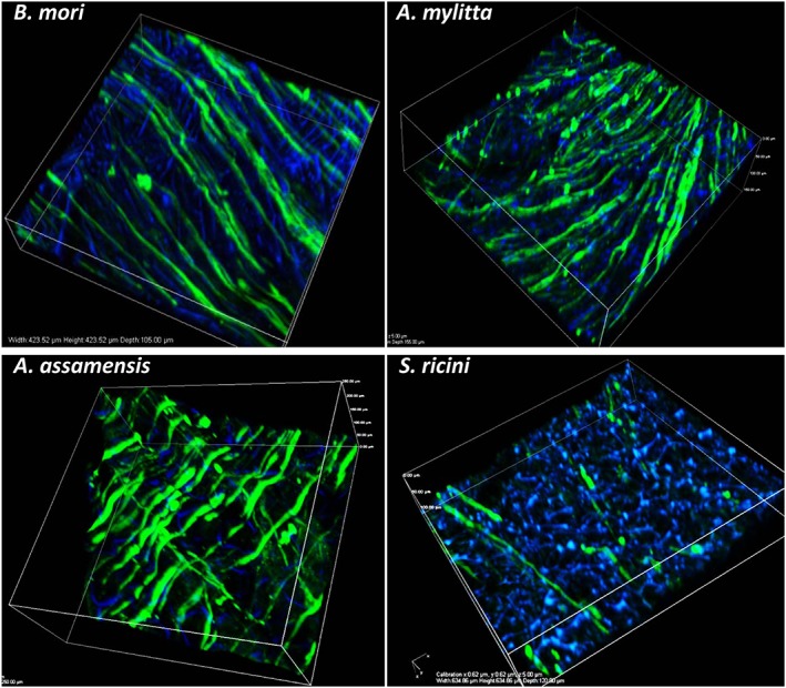
Figure 3.

Immunostained images of differentiated human skeletal muscle myoblasts (HSMMs) on three‐dimensional (3D) silk scaffolds. The HSMMs were cultured on the silk scaffolds for 4 days in proliferation medium and a further 4 days in differentiation medium. After differentiation, the scaffolds were stained with anti‐myosin monoclonal antibody (NOQ7.5.4D) and goat anti‐mouse IgG‐AF488. The nuclei are stained with 4′,6‐diamidino‐2‐phenylindole (DAPI, blue). Images were captured using a Nikon A1 confocal microscope. The 3D merged images of several z‐stack images (5 μm each stack, 150–200 μm deep into scaffold) are represented. Bar: 100 μm. [Colour figure can be viewed at wileyonlinelibrary.com]
