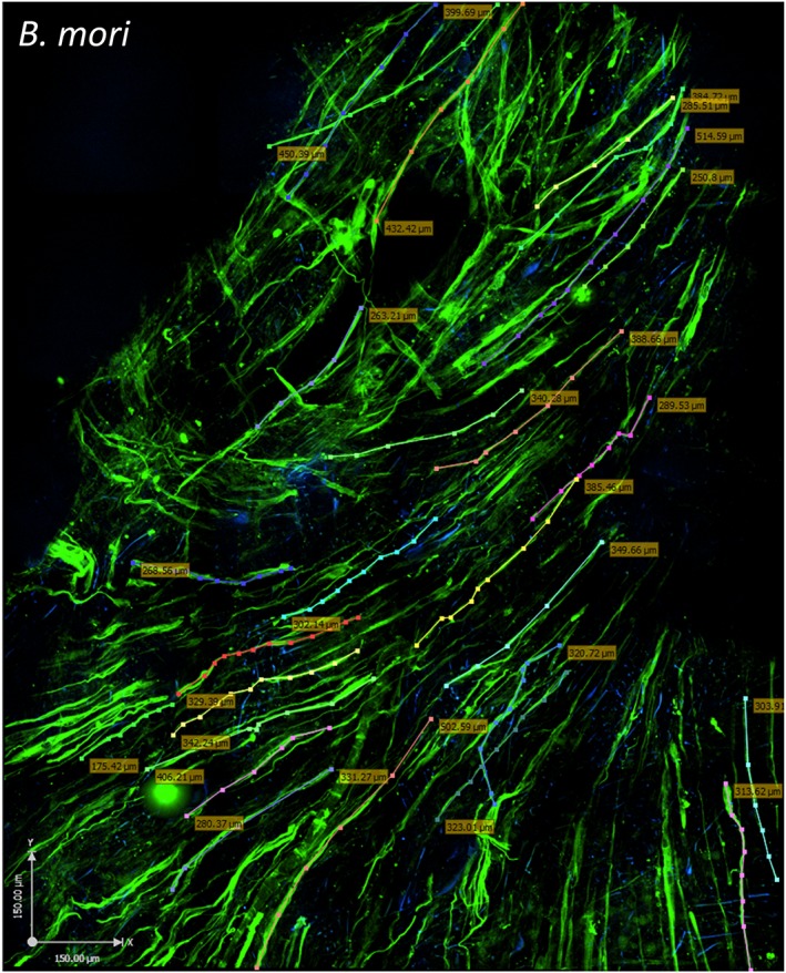
Figure 4.

Human skeletal muscle myoblasts (HSMMs) form a three‐dimensional (3D) muscle‐like tissue on Bombyx mori scaffolds. The HSMMs were cultured for 4 days in skeletal muscle growth medium‐2 proliferation medium and 4 days in differentiation medium on B. mori silk scaffolds. Myotubes were stained with anti‐myosin monoclonal antibody (NOQ.5.4.D), followed by a goat anti‐mouse AF488 conjugated second antibody. The images were captured using an Ultraview spinning disc confocal microscope (PerkinElmer). To capture a large area and estimate the coverage of the myotubes on the scaffold surface, 50 z‐stack images (2.5 μm each stack) were stitched together using Volocity software. Muscle fibre length (coloured dotted lines and boxed numbers) was measured by tracing individual fibres using Volocity software. The area covered in this image is indicated by the following: x‐axis, 1.58 mm; y‐axis, 1.213 mm; z‐stack, 125 μm. Bar: 150 μm. [Colour figure can be viewed at wileyonlinelibrary.com]
