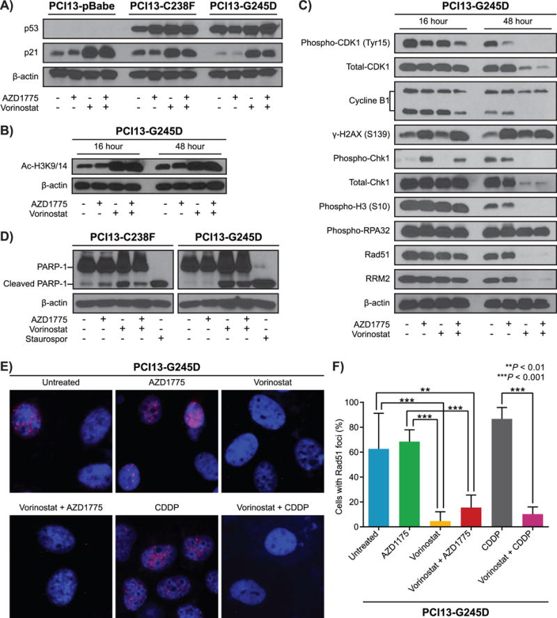
Figure 4. The synergism between vorinostat and AZD1775 in HNSCC mutp53 cells is mediated by increased replication stress associated with impaired Rad51-mediated homologous recombination and apoptosis.

PCI13-pBabe and PCI13 with mutant TP53 (C238F, G245D) cells treated with either vorinostat, AZD1775 alone or in combination for 16 and/or 48 hours and subjected to immunoblot analysis using antibodies as indicated. A and B, protein expression levels of p53, p21 and acetylated histone 3 respectively. C, levels of phosphorylation of H2AX (S139), Chk1 (S345), total Chk1, hyperphosphorylation of RPA32, Rad51, and RRM2. The mitotic entry marker histone H3 S10 phosphorylation, cyclin B1 total protein, CDK1 (CDC2) tyr15 phosphorylation, and total protein levels were also analyzed. D, western blots for HNSCC cells with mutant TP53 (PCI13-C238F, and PCI13-G245D) treated with either vorinostat, AZD1775 alone or in combination as indicated and analyzed for the presence of PARP-1 cleavage as marker of apoptosis. Lysates from staurosporine-treated (1 μmol/L) cells were used as positive controls for apoptosis. β-actin served as loading control .E, vorinostat treatment lead to reduced Rad51 focus formation, indicating impaired Rad51-mediated homologous recombination in HNSCC cells with high-risk TP53 mutation. F, quantification of Rad51 foci images shown in Figure 4E.
