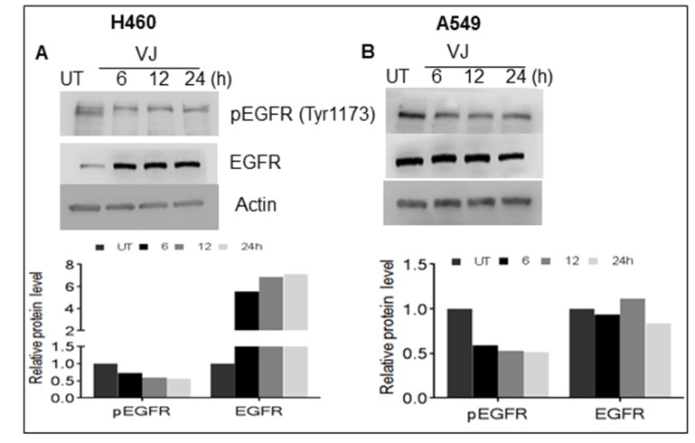
Figure 4. Cells were treated with VJ at IC50 concentrations for 6, 12, 24 hrs. and cell lysates quantified for western blot analysis for effect on basal level of EGFR and phosphorylated EGFR in A. H460 and B. A549 cells.
β-Actin was used as loading control. The densitometry analyses of bands are expressed in arbitrary units. Analysis was performed using Image Studio Lite 5.2 software.

