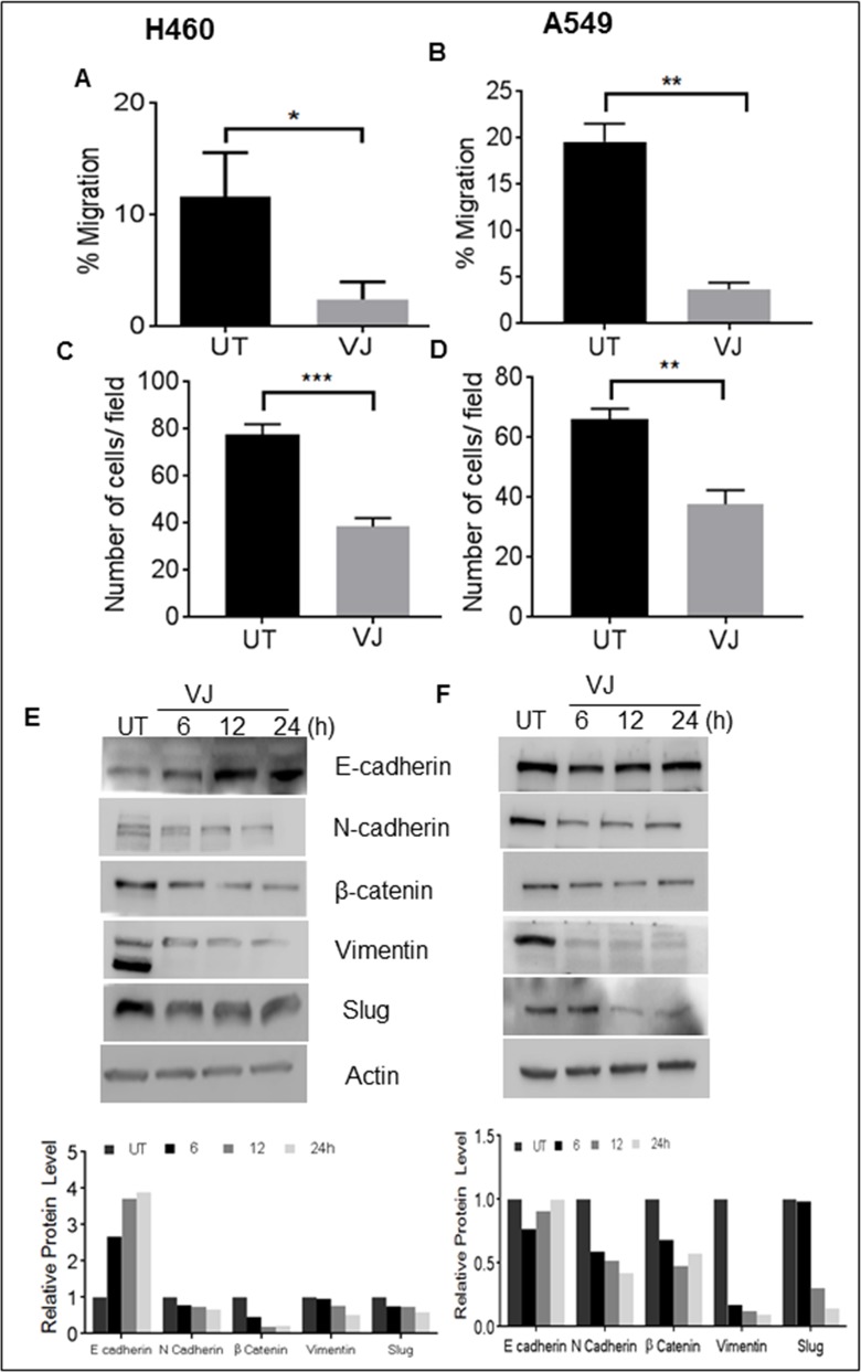
Figure 7. VJ inhibits EMT in H460and A549 lung cancer cells.
A., B. Wound healing assay: a wound was created and the H460 and A549 cells were treated with resp. The wound gap was photographed at the same points using the distance between the two edges of wound (ImageJ). C., D. Transwell invasion assay performed using Boyden chambers. The H460 and A549 invaded cells were stained with crystal violet and counted. E., F. The H460 and A549 cells were treated with VJ and DMSO (as control), lysates were prepared after 6, 12, 24 hr treatment period and analyzed for E-cadherin, N-cadherin, β-catenin, Vimentin, slug proteins with western blotting. β-Actin was used as loading control and the densitometry analysis of bands are expressed in arbitrary units. Analysis was performed using Image Studio Lite 5.2 software. Student's t-test was used to calculate statistical significance between VJ treated and untreated at each time point *p < −0.05, **p < 0.01, ***p < 0.001.

