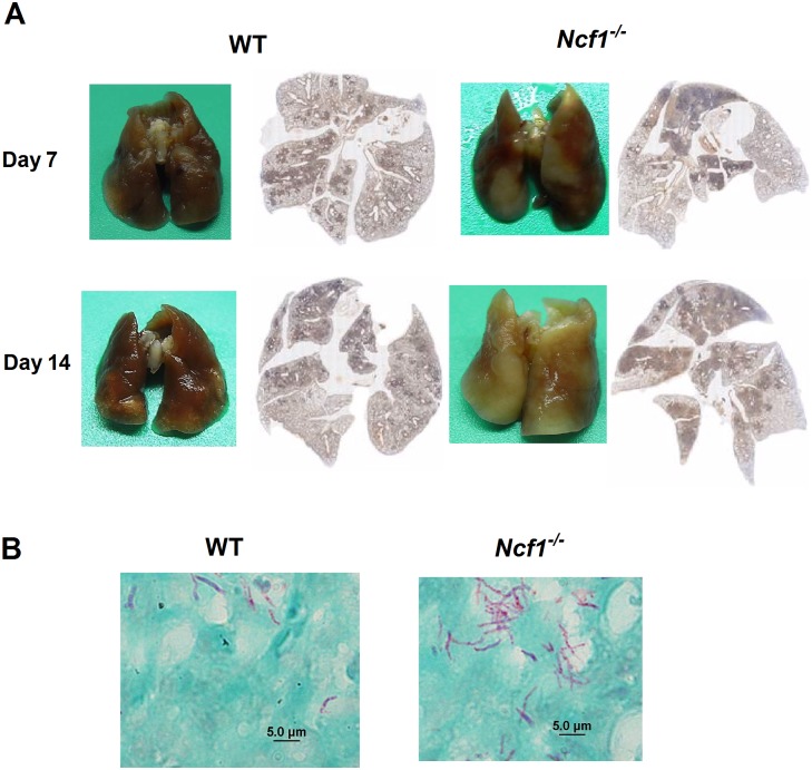
Fig 2. A Higher level of pulmonary inflammation in Ncf1-/- mice after M. marinum infection in comparison with the inflammation in WT mice.
Representative gross pictures (A) and cross-sectional histological examinations (B) of M. marinum-infected lungs from WT and Ncf1-/- mice at day 7 and day 14 after infection. Acid-fast stains (AFS) (B) at day14 showed abundant AFS-positive bacilli in Ncf1-/- mice, whereas sparse AFS-positive bacilli were found in WT mice. These experiments were repeated with similar results.

