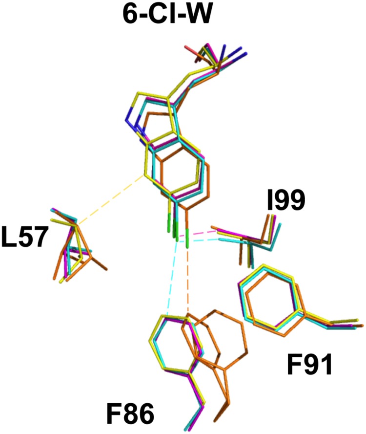
Fig 6. Structural overlay of M011, 78A, P2 peptides bound to Mdm2.
Only the tryptophan residue of each peptide (with 6-chloro group shown in green) and amino acids forming the Mdm2 Trp pocket depicted. M011: cyan, magenta; 78A: orange; P2: yellow. Corresponding Mdm2 residues for each peptide are in the same colour. Image generated using structures 2GV2, 2AXI and 5XXK. Dotted lines represent the shortest Van der Waals contacts. Formulae for the complete P2 and 78A peptides are N-acetyl-Phe-Met—Phe(4-MePhos)-Trp(6-Cl)-Glu-(AC3C)-Leu amide and cyclo(-Phe-Glu-(6-Cl)Trp-Leu-Asp-Trp-Glu-Phe-d-Pro-Pro-), respectively.

