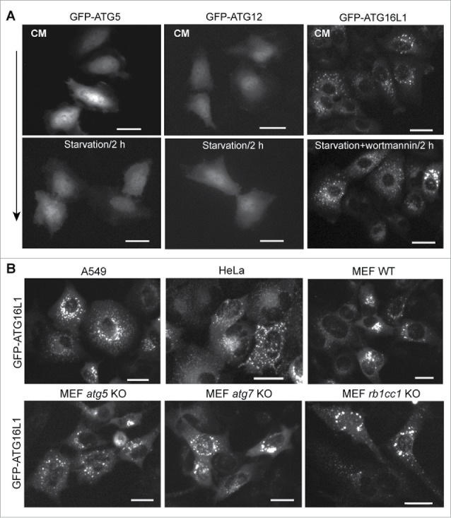
Figure 2.

Puncta formation of transiently expressed ATG16L1 is independent of PtdIns3K activity and the ATG12–ATG5 conjugate. (A) HeLa cells were cultured in a 6-well plate at 2 × 10⁵ cells per well for 16 h. Cells were then transfected with 500 ng of GFP-ATG5, GFP-ATG12, or GFP-ATG16L1 expression plasmid per 6-well plate for 20 h, respectively. Cells were starved in EBSS with/without wortmannin (200 nM) for 2 h. (B) A549 cells, HeLa cells, MEF WT, MEF atg5 KO, MEF atg7 KO, or MEF rb1cc1 KO cells were transduced with adenoviral vector expressing GFP-ATG16L1 at 2 MOI for 20 h, respectively. Scale bar: 25 micron.
