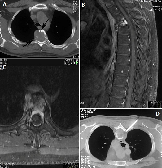
Figure 1.

(A) CT scan depicts an expansile lytic lesion in T6 vertebra that destroys the cortex of the vertebral body (arrows), Sagittal; (B) and axial; (C) fat-saturated (FS) T1-weighted MRI with intravenous gadolinium contrast demonstrates compression fracture of vertebral body and the extent of disease in the marrow cavity, soft tissues and epidural space; (D) computed tomography guided needle biopsy
