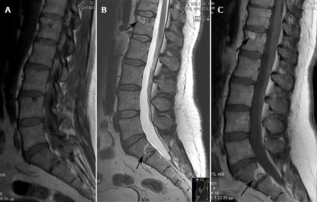
Figure 3.

MRI of the lumbar spine, sagittal section, T1: (A) T2; (B) and gadolinium-enhanced T1-weighted; (C) sequences: presence of nodular bone metastases with a halo (arrows)

MRI of the lumbar spine, sagittal section, T1: (A) T2; (B) and gadolinium-enhanced T1-weighted; (C) sequences: presence of nodular bone metastases with a halo (arrows)