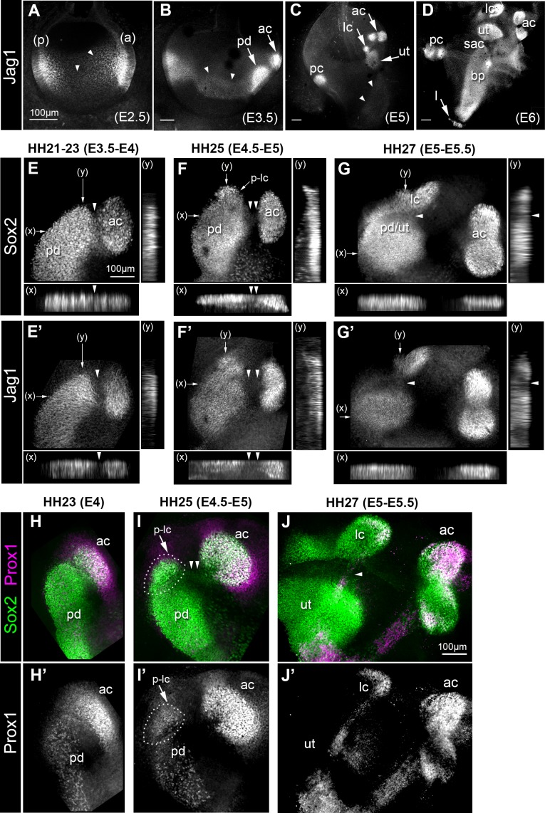
Figure 1. Expression of the Notch ligand Jag1,Sox2 and Prox1 during the formation and segregation of the chick inner ear sensory patches.
(A–D) Jag1 expression in the developing chick inner ear (whole-mount preparations). At E2.5 (A), two patches of high expression are present in the anterior (a) and posterior (p) poles of the otocyst, but lower levels of expression are also detected in the medial region along the antero-posterior axis (arrowheads). At E3.5 (B), two patches of high Jag1 expression are now present in the anterior domain (arrows), corresponding to the anterior crista (ac) and a larger prosensory domain (pd); the medial domain of lower expression remains clearly visible (arrowheads). At E5 (C), strong Jag1 staining is observed in the posterior (pc), anterior and lateral (lc) cristae and the utricle (ut); another fainter domain of expression extends ventrally within the developing cochlear duct (arrowheads). At E6 (D), the utricle, the three cristae, and the lagena (l) are well defined. Jag1 expression still forms a continuous domain of expression between the saccule (sac) and the basilar papilla (bp). (E–G’) Whole-mount surface views and transverse projections (along the x and y directions) of the anterior prosensory domains at different developmental stages, immunostained for Sox2 and Jag1 expression. At HH21-23 (E–E’), the prospective anterior crista becomes apparent at the edge of a large prosensory domain (pd); a group of Sox2-positive and Jag1-negative cells (arrowheads) is located at the interface of the two prosensory patches. At HH25 (F–F’), the distance separating the anterior crista from the large prosensory domain increases (double arrowheads); a distinct cluster of Sox2-positive cells becomes visible at the upper edge of the large prosensory domain, which corresponds to the location of the prospective lateral crista (p–lc). At HH27 (G–G’), the lateral crista starts to segregate from the prospective utricle (pd/ut); the interface domain is Sox2-positive, but exhibits a marked down-regulation of Jag1 expression (arrowheads). At HH23 (H–H’), Prox1 is strongly expressed in the prospective anterior crista (ac), which is still connected to the large prosensory domain (pd). At HH25 (I–I’), strong Prox1 immunostaining is present in the anterior crista; a new, denser cluster of Prox1-positive nuclei (dotted outline) is also present at the superior edge of the large prosensory domain, presumably corresponding to the prospective lateral crista (p–lc). Note that the cells located in between the anterior crista and the large prosensory domain still retain faint Sox2 expression (arrowheads in I). At HH27 (J–J’), the lateral crista (lc) segregates; note that only one of its halves contains Prox1-expressing cells at this stage. Scale bar for all panels = 100 μm.


