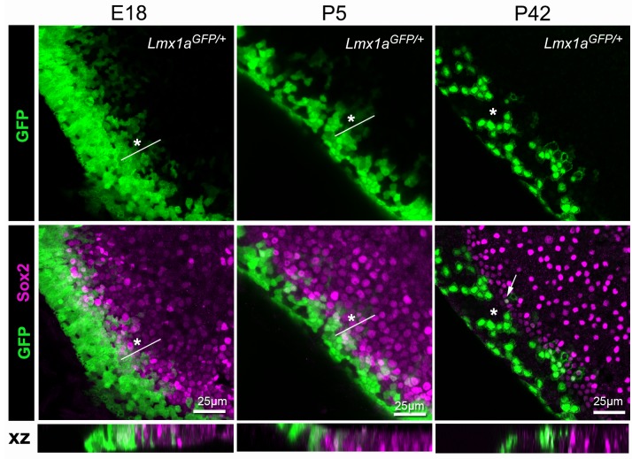
Figure 11. Lmx1a expression is progressively down-regulated at the lateral border of the developing utricle.
Maximum intensity z-projections of whole-mount utricles from Lmx1aGFP/+ mice at different developmental ages (E18, P5 and P42), immunostained for Sox2 expression (magenta). Note the mosaic pattern of GFP fluorescence at the lateral border of the utricle, and the overlap between GFP+ and Sox2+ cells at E18 and P5 (asterisk). In the adult (P42) utricle, only a few cells retained low levels of GFP fluorescence and Sox2 expression (arrow) in the sensory domain, whilst mosaic expression of GFP is still observed in the transitional cell region (asterisk). The XY images are orthogonal projections taken from the same preparations.

