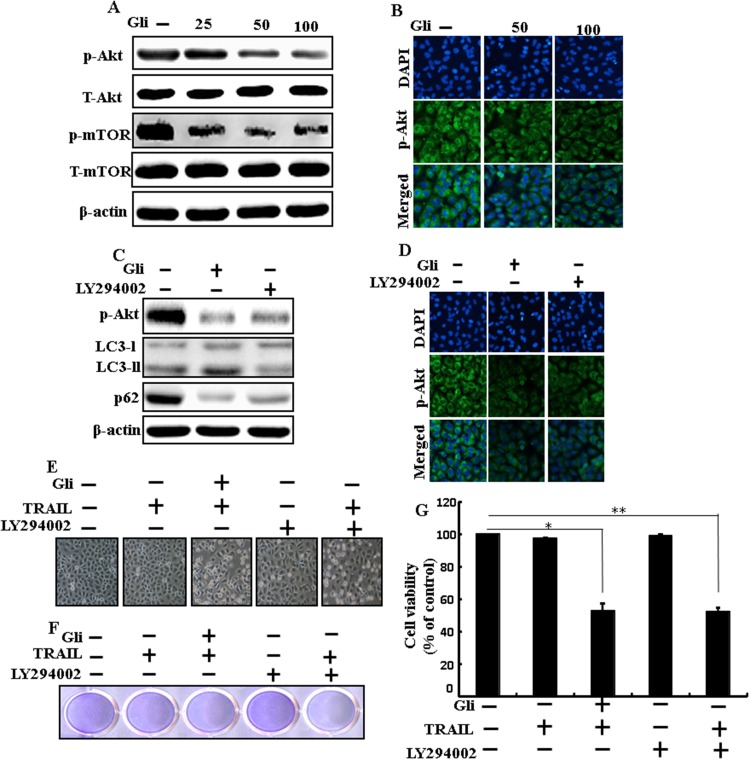
Figure 7. Effects of glipizide on the Akt/mTOR/autophagy signaling pathway.
Lung adenocarcinoma cells were pre-incubated with different doses of glipizide (0, 25, 50, and 100 μM) for 12 h and exposed to TRAIL protein for an additional 2h. Additional cells were pretreated with LY294002 for 1 h followed by treatment with glipizide. After that, (A and C) western blot for T-Akt, p-Akt, p-mTOR, T-mTOR, LC3-II, and p62 proteins was analyzed from A549 cells; (B and D) Cells were immunostained with p-Akt antibody (green) and observed in fluorescent view; (E) Cell morphology photographed using light microscope (×100); (F) Cell viability was measured with crystal violet assay; (G) Bar graph indicating average density of crystal violet. β-actin was used as loading control. *p < 0.05, **p < 0.01: represent significant differences between control and each treatment group; Gli: Glipizide; TRAIL: Tumor necrosis factor (TNF)-related apoptosis-inducing ligand.

