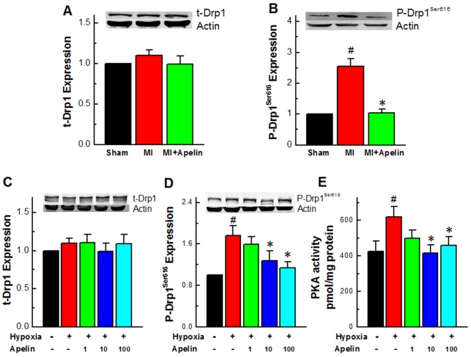
Figure 2. Apelin reduces the activity of Drp1.
(A) Expression levels of Drp1 in MI mice. (B) Expression levels of p-Drp1Ser616 in MI mice. (C) Expression levels of Drp1 in cultured neonatal mice cardiomyocytes. (D) Representative western blot band of p-Drp1Ser616 in cultured neonatal mice cardiomyocytes. (E) PKA activity in primary cultured cardiomyocytes under a hypoxic condition. Data are presented as mean ± SEM, #P < 0.05 vs sham, n = 6; *P < 0.05 vs MI, n = 8. P values were analyzed using one-way ANOVA.

