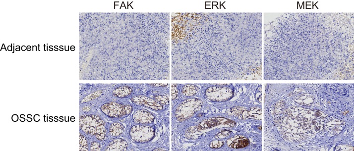
Figure 9. FAK, ERK and MEK expressions in the OSCC tissues and the normal adjacent tissues (× 200).
FAK and MEK were mainly expressed as pervasive and brown particles in the cytoplasm, while ERK was expressed primarily in the nucleus as indicated by brown staining. The positive expression rates of these three proteins in the OSCC tissues were all significantly higher than those in the adjacent normal tissues; OSCC, oral squamous cell carcinoma; FAK, focal adhesion kinase.

