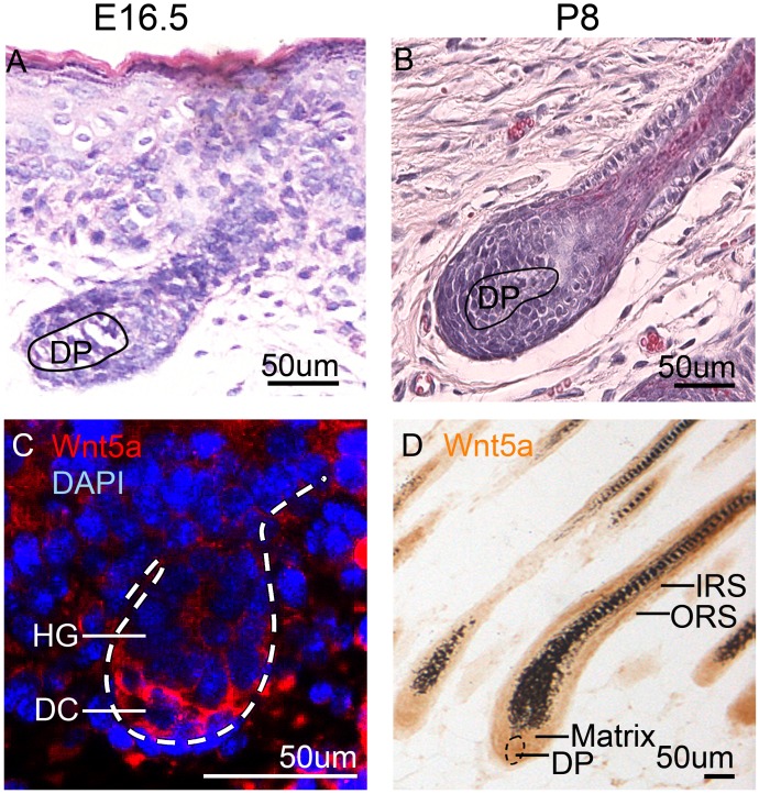
Figure 1. Wnt5a expression during hair development and growth.
(A) H&E staining shows a developing hair follicle at E16.5. (B) H&E staining shows a developed hair follicle in full anagen at P8. (C) Immunofluorescent staining shows that Wnt5a is expressed in the mesenchymal cells of skin, particularly in the DP at E16.5. (D) Immunohistochemistry staining shows that Wnt5a is expressed in the IRS, ORS, hair matrix and DP cells. E16.5, embryonic day 16.5; P8, postnatal day 8; DC, dermal condensation; DP, dermal papilla; HG, hair germ; IRS, inner root sheath; ORS, outer root sheath. N=9.

