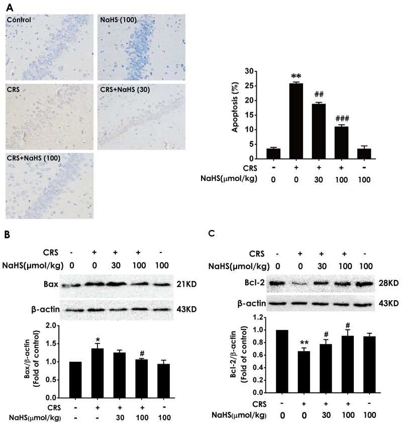
Figure 4. Effect of H2S on CRS-induced hippocampal apoptosis in rats.
1 day after behavior tests, the hippocampus of rats were collected. (A) The level of apoptosis was detected by tunel staining (Left, magnification x400). (B and C) The apoptotic-associated proteins Bax (B) and Bcl-2 (C) were measured by western blotting. Values are expressed as the mean ±S.E.M. (n=3 per group). *P < 0.05, **P < 0.01, vs control group; #P < 0.05, ##P < 0.01,###P < 0.001, vs CRS-treated alone group.

