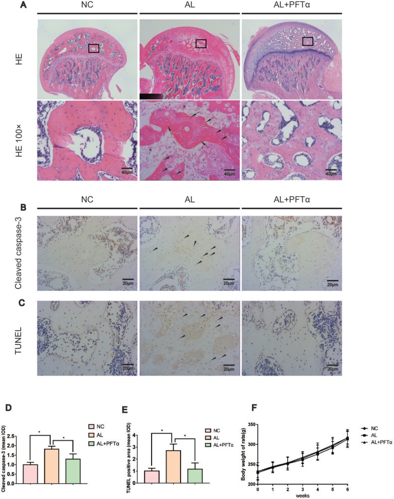
Figure 5. The development of alcohol-induced ONFH in the rat model.
(A) H&E staining of the femoral head indicated obvious signs of osteonecrosis in the AL group. Empty lacunae or pyknotic nuclei (black arrows) were present in the subchondral trabeculae of the AL group, and the accumulation of bone marrow hematopoietic cellular debris was present in the medullary space (black stars). Significantly less ONFH change was detected in the AL+PFTα group. (B) Immunostaining of cleaved caspase-3 in the femoral heads is shown. Positive staining was present in the trabeculae of the femoral head in the AL group (black triangles). (C) TUNEL staining of the femoral head is shown. Positive staining was present in the trabeculae of the femoral head in the AL group (black triangles). (D–E) Quantification of positive staining of cleaved caspase-3 and TUNEL. (N = 3, *significant difference versus the AL group, *p < 0.05) (F) No significant difference in body weight was observed among the three rat groups.

