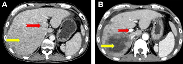
Figure 3. Image of a 48-year-old man with hepatocellular carcinoma and PVTT who showed a partial response after combined apatinib and TACE treatment.
Contrast-enhanced CT at diagnosis showed a 134 mm diameter hepatocellular carcinoma nodule (yellow arrow) and multiple small metastatic lesions located in the liver, together with PVTT in the left and main portal vein (red arrow). CT images 1 month after diagnosis showed intrahepatic lesions in numerous non-enhanced areas (yellow arrow) and almost complete absence of PVTT without definite enhancement (red arrow).

