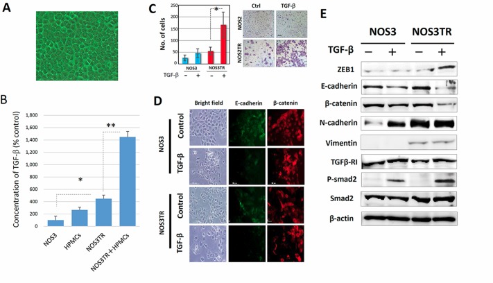
Figure 5. Enhanced ZEB1 expression by microenvironmental production of TGF-β.
(A) The appearance of the cultured human peritoneal mesothelial cells (HPMCs). HPMCs typically showed an epithelial morphology with a cobblestone appearance. (B) TGF-β concentrations in conditioned media of HPMCs, NOS3, NOS3TR, and HPMCs plus NOS3TR cells (co-culture). TGF-β production in NOS3TR cells was significantly higher than in parental NOS3 cells. Asterisks show significance (P < 0.05). (C) The effect of TGF-β on migratory potential of parental or PTX-resistant cells. The addition of TGF-β to NOS3TR cells led to a significantly enhanced migratory potential compared with parental NOS3 cells (Right panel). Asterisks show significance (P < 0.05). Left panel: representative images. (D) The immunofluorescence expressions of E-cadherin and β-catenin in NOS3 or NOS3TR cells with or without TGF-β. The presence of TGF-β caused a more mesenchymal cell shape with the loss of cell polarity and decreased E-cadherin expression, particularly in NOS3TR cells. (E) Western blot analysis of the EMT, or TGF-β signal-related proteins in NOS3 and NOS3TR cells with or without TGF-β. The addition of TGF-β resulted in increased ZEB1 expression with the enhanced expression of phosphorylated Smad 2, particularly in NOS3TR cells.

