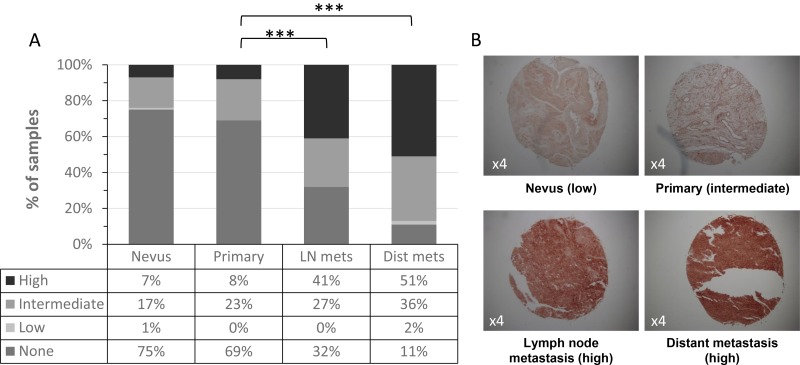
Figure 2. ICAM-1 expression with melanoma development and progression.
(A) ICAM-1 membrane expression was analyzed in melanoma progression TMA comprised of nevi, primary tumors, lymph node (LN) metastases and distant metastases. Intensity staining of ICAM-1 was scored as none, low, intermediate or high; (B) Representative staining patterns (×4) of ICAM-1 in melanocytic specimens.

