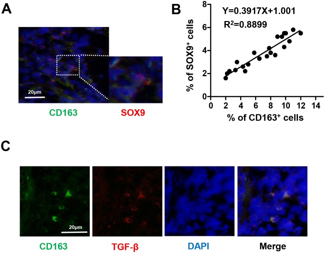
Figure 1. Number of TAMs is correlated with SOX9 expression in NSCLC.
(A) Immunofluorescent staining of human NSCLC tumor tissues. CD163+ macrophages are stained green and SOX9+ lung cancer cells are stained red. Nuclei were stained with DAPI (blue). (B) Linear regression revealed a positive correlation between numbers of CD163+ macrophages and SOX9+ lung cancer cells. CD163+ macrophages and SOX9+ lung cancer cells within a distance of 100 μm were counted in 22 separate microscope fields. (C) CD163+ macrophages express TGF-β; CD163 is stained green, TGF-β red, and nuclei blue.

